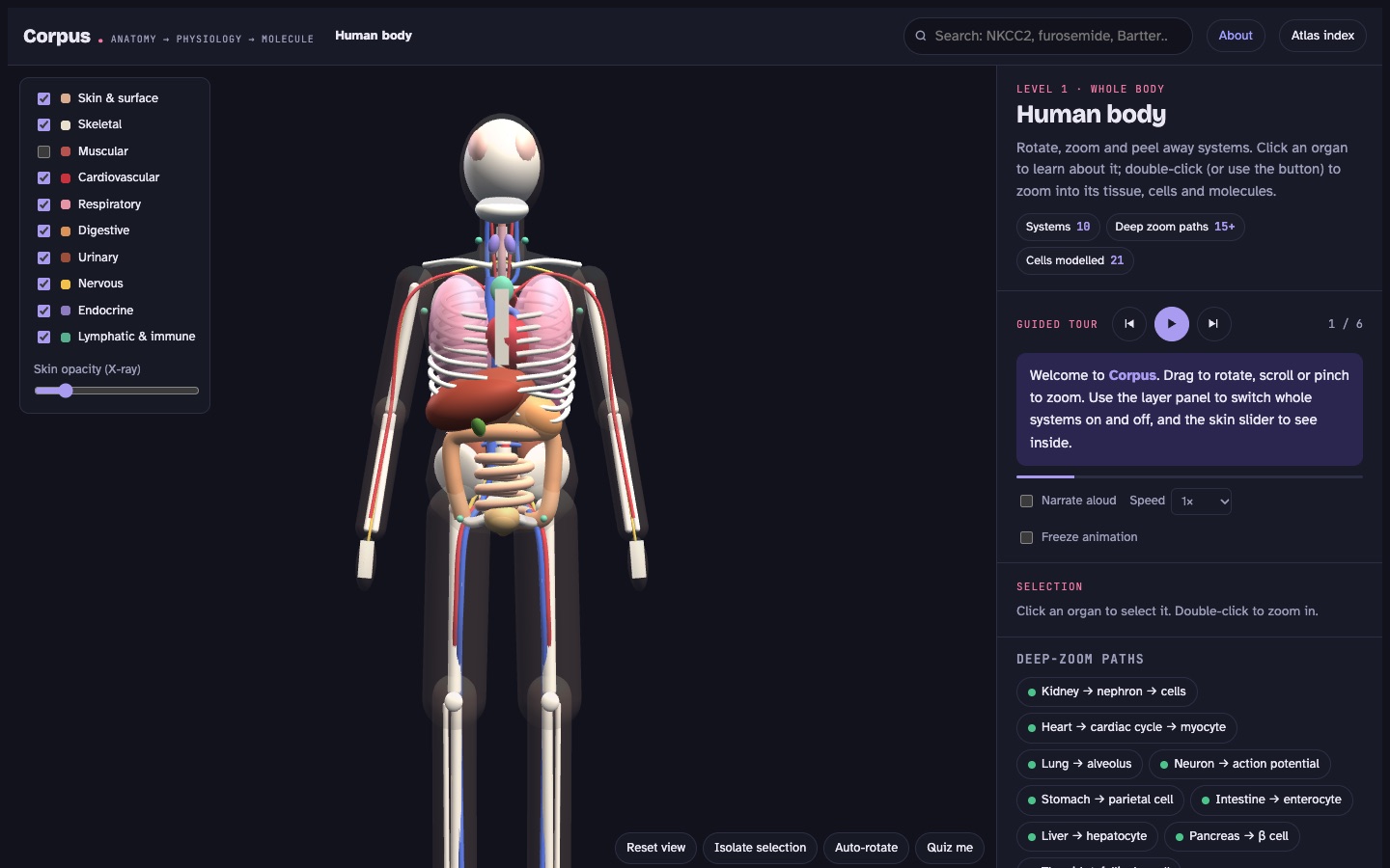
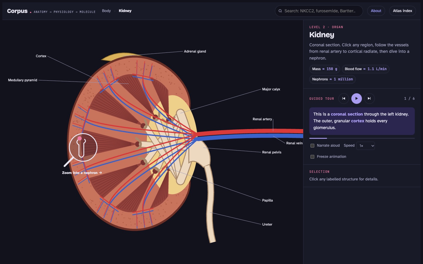
Most 3D anatomy apps stop at the organ. Corpus keeps going: into the nephron, onto the membrane of one cell, down to each transporter, with its gene, what it carries, the hormones that tune it, the drugs that block it and the diseases that break it.
Runs in your browser. No account, no tracking, works offline once installed.

Every level is clickable and every structure explains itself. Breadcrumbs keep you oriented, and search jumps straight to any gene, drug or disease.


The heart beats, filtrate concentrates, oxygen loads and nerves fire. Move a slider and the whole system responds, from urine output to cardiac output.
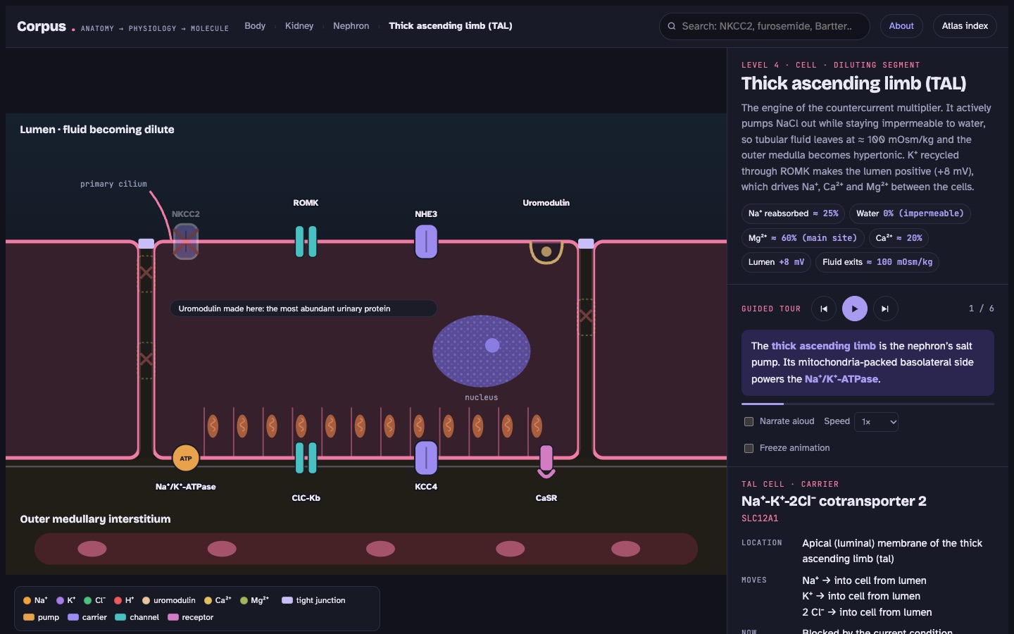
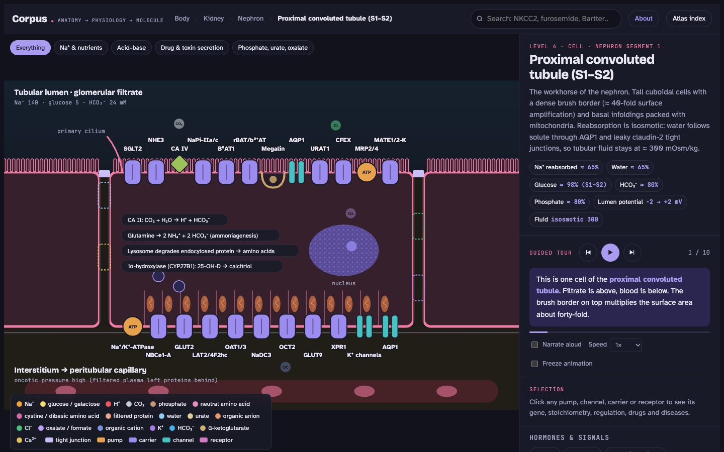
Each pump, channel, carrier and receptor sits on the membrane where it is actually expressed, moving the right ions in the right ratio.
Turn on furosemide, ADH, Bartter syndrome or cystic fibrosis and watch which transporters stop, speed up or appear, with the clinical consequences explained.
Every level has a step-by-step tour that highlights each structure while it explains it, read aloud by a natural recorded voice with the current sentence highlighted.
Switch any organ, tissue or cell to Histology and get a real stained micrograph with numbered pins on the actual structures, pan and zoom, a guided tour and a quiz. A drawing mode shows a simplified guide.
Find the spleen on the 3D body, or locate NKCC2 on a cell with every label hidden. Score yourself as you go.
Type a gene (SLC12A3), a drug (amiloride) or a disease (Liddle) and land on the exact transporter, with the condition already applied.
Each one is a quantitative teaching model with clinical presets.

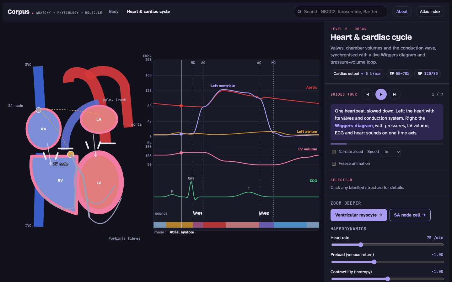
Valves, chamber volumes and the conduction wave, synchronised with a Wiggers diagram and pressure–volume loop.

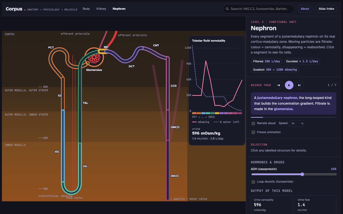
A countercurrent model of a juxtamedullary nephron. Tubular fluid changes colour with osmolality and thins out as it is reabsorbed.

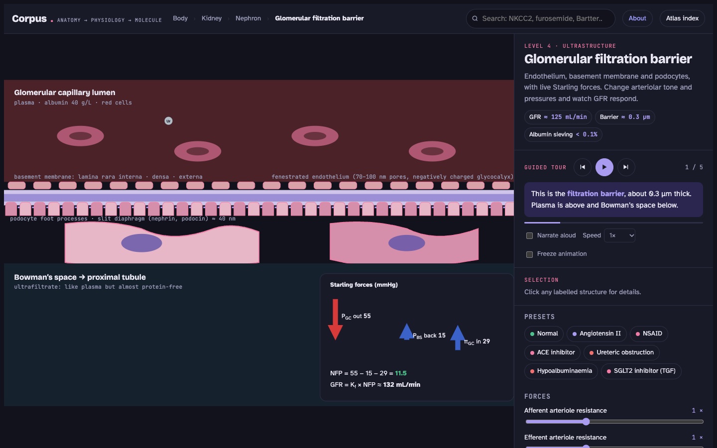
Endothelium, basement membrane and podocytes with live Starling forces.

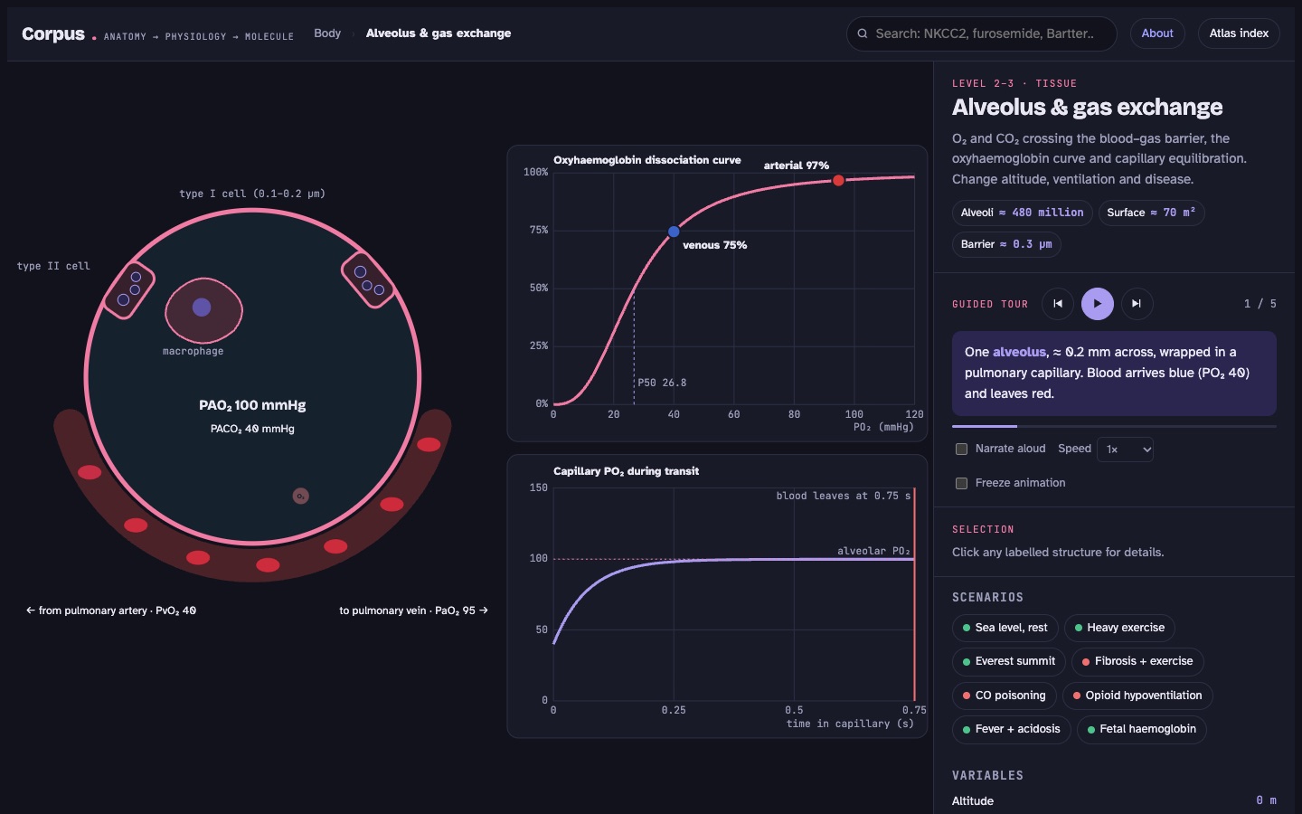
An alveolus with oxygen and CO₂ crossing the barrier, the oxyhaemoglobin curve and capillary equilibration.

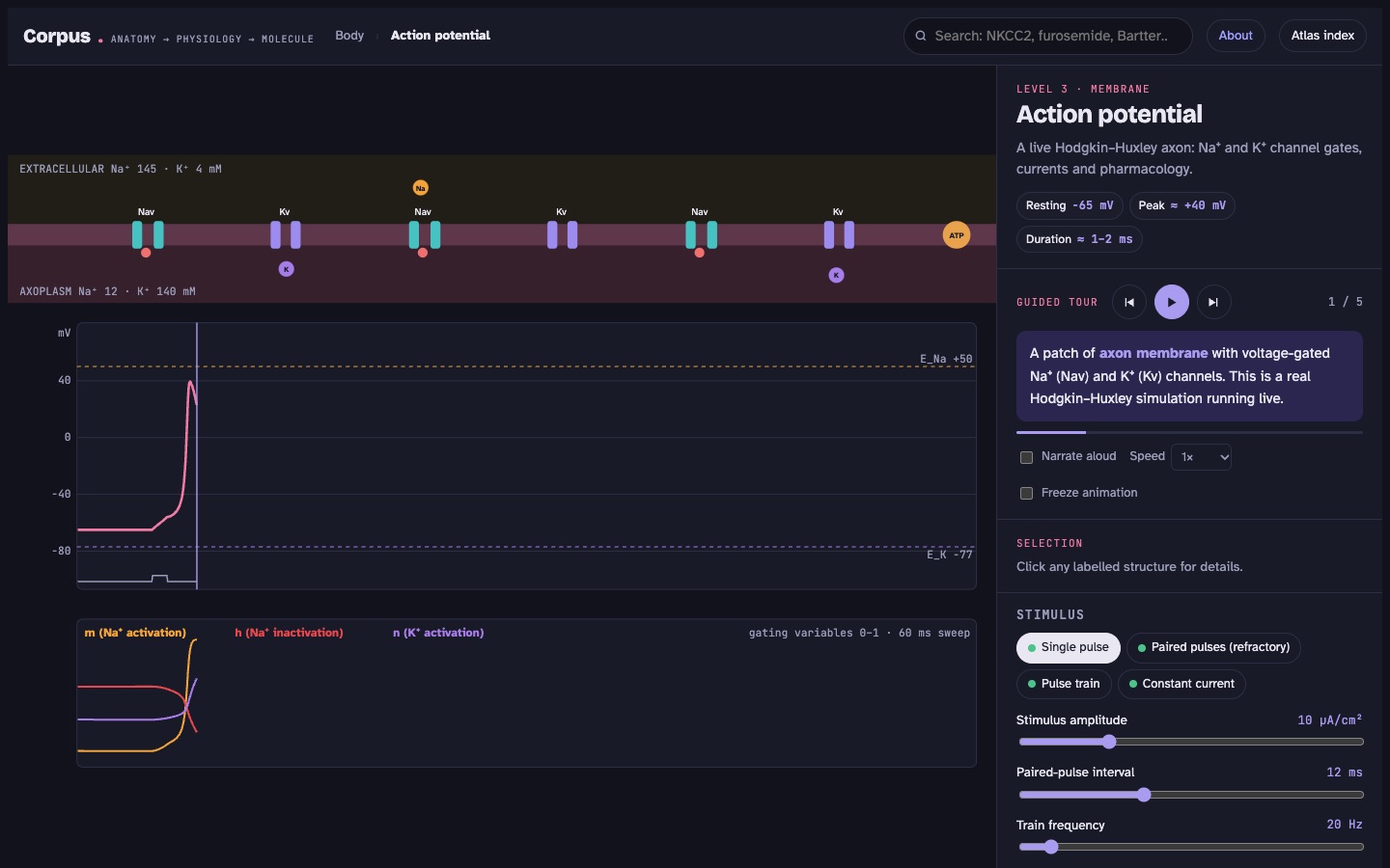
The Hodgkin–Huxley equations integrated live in your browser, with channel gates you can watch open and close.

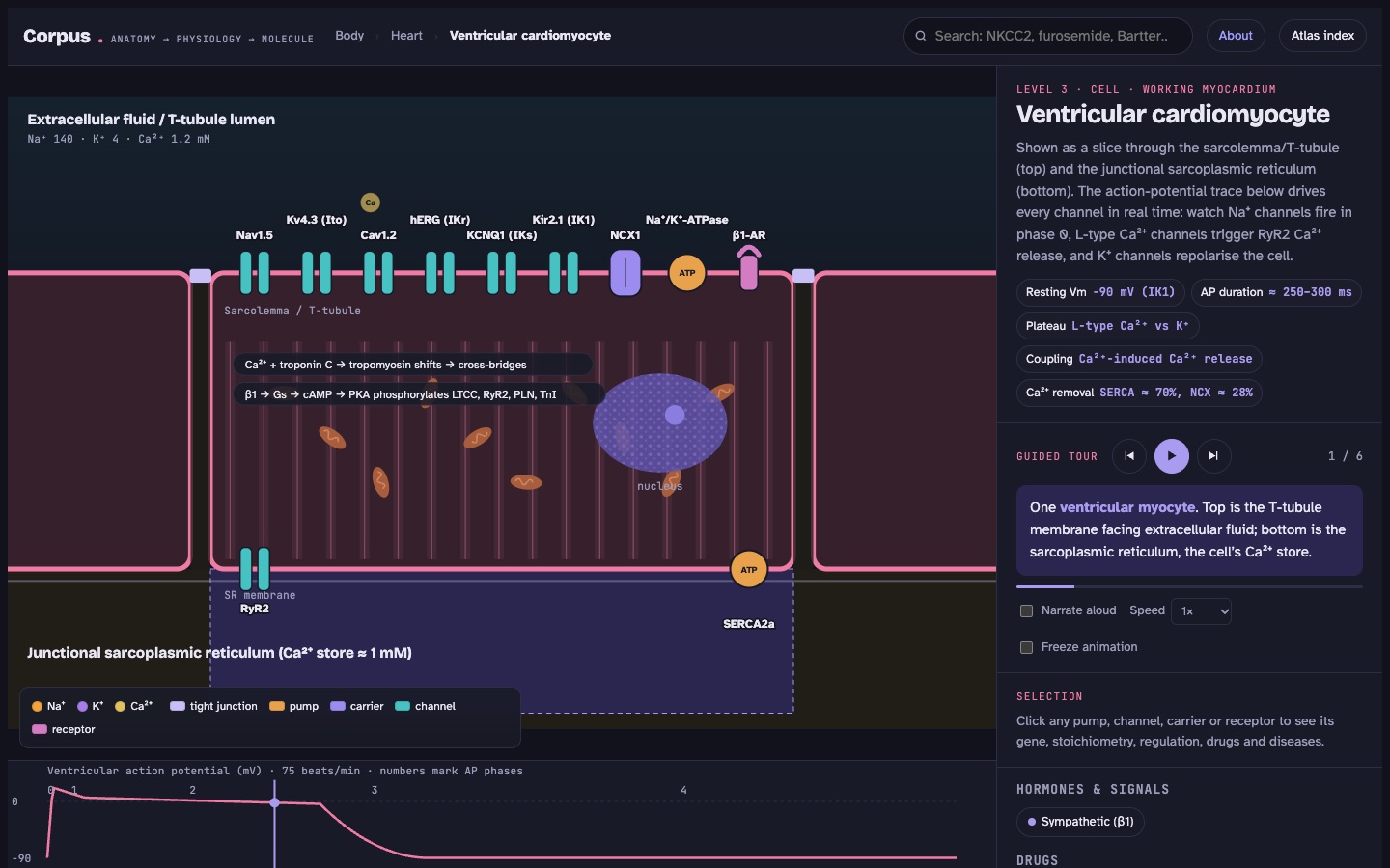
In the myocyte and pacemaker cell, an action-potential trace drives every channel in real time.
Stomach → gastric gland → parietal cell. Intestine → villus → enterocyte. Liver → lobule → hepatocyte. Pancreas → lobule → β cell. Thyroid → follicle → thyrocyte. Lungs → acinus → alveolus. Brain → neuron → action potential. Heart → heart wall → myocyte. Colon → crypts → colonocyte. Bladder → wall → detrusor cell. Adrenal → zones → glomerulosa cell. Femur → osteon → osteoclast. Biceps → sarcomere (contracting live) → muscle fibre. Skin → epidermis and sweat duct. Arteries → artery wall → smooth-muscle cell. Plus the spleen, oesophagus, pituitary, thymus and lymph node, each down to its tissue. Every animation can be slowed to 0.1× or paused.
Proximal tubule S1–S2 and S3, thin descending and ascending limbs, thick ascending limb, macula densa, DCT, principal cell, α- and β-intercalated cells, inner medullary collecting duct. Plus the organ, nephron and glomerular barrier.
Ventricular cardiomyocyte and sinoatrial pacemaker cell, plus the cardiac cycle with conduction system and valves.
Ciliated airway epithelial cell (CFTR, ENaC), plus the alveolus with type I and II pneumocytes and macrophages.
Gastric parietal cell, villus enterocyte, crypt secretory cell and hepatocyte (bile secretion).
Pancreatic β cell (insulin secretion), pancreatic duct cell (HCO₃⁻) and thyroid follicular cell.
Skeletal muscle fibre (end-plate and triad, with myasthenia, malignant hyperthermia and neuromuscular blockers), detrusor and vascular smooth muscle, osteoclast (osteoporosis drugs), and sweat duct cell (the cystic fibrosis sweat test).
Surface colonocyte (aldosterone, K⁺ secretion, butyrate, secretory diarrhoea) and zona glomerulosa cell (aldosterone synthesis and adenoma mutations).
Axon membrane with Nav and Kv channels in a live Hodgkin–Huxley model, and peripheral nerves, cord and brain in 3D.
Corpus is a set of static files with no server and no account. Pick whichever way suits you.
In Chrome or Edge, use the Install button or the install icon in the address bar. On iPhone or iPad, tap Share → Add to Home Screen. It then opens in its own window and works offline.
A self-contained offline copy. Unzip it and double-click Corpus.html. No installation and no internet needed.
Download Corpus.zipOptional: Corpus-voices.zip adds the recorded narration. Unzip it into the same folder.
Transporter locations, stoichiometries, genes, regulation and pharmacology follow standard physiology and pharmacology teaching. The quantitative models (urine concentration, Starling forces, the Wiggers diagram, gas exchange) are simplified teaching models that reproduce textbook values, not patient simulators.
It is BodyParts3D, an adult male reference anatomy built from MRI data and refined by anatomists (© Database Center for Life Science, CC BY 4.0), with 2,234 bones, muscles, vessels, nerves and organs you can select one by one. It is one reference body, so it does not show every variation, and a few structures (lung tissue, the thyroid gland, lymph nodes) are not in the dataset. The offline download uses a lighter schematic body instead.
Medical, nursing, pharmacy and physiology students, teachers who want something to project, and anyone curious about how the body works at the level of molecules.
No. Corpus is for learning. It does not give medical advice and should not guide diagnosis or treatment.